


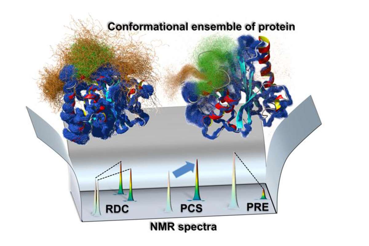
Ensemble map of protein structure.The team’s approach integrates different NMR data to create a map of how different parts of a molecular structure move. Credit: Tokyo Metropolitan University.
A new Nuclear Magnetic Resonance (NMR) method has revealed atomic scale dynamics play a role in yeast enzyme function.
Researchers from Tokyo Metropolitan University have developed a new structure determination method using NMR spectroscopy which shows how different parts of complex molecular machinery like enzymes move while they help catalyse reactions.
Focusing on an enzyme in yeast, they demonstrated how contrasts in atomic scale motions impact their function.
The method promises unprecedented access to the mechanisms by which biomolecules work, and how they relate to illnesses.
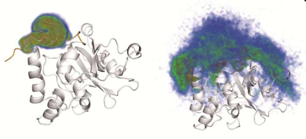
Probability density map for location of crossover loop and gating lid.Around the protein-capturing component and the main body of the protein, shown in white, the crossover loop (left) and the gating lid (right) are in constant motion such that they can be found in a wide range of locations, as illustrated by the coloured clouds. Credit: Tokyo Metropolitan University. CC BY-NC-ND.
Blur of motion
Enzymes are indispensable to the function of all biological organisms, including humans. While snapshots captured using X-ray or cryo-electron microscopy have revealed their intricate molecular structure, they are a constant blur of motion when they are at work.
Their atomic scale structure is constantly changing, capturing other biomolecules and helping them react in a carefully choreographed sequence.
While clearly important, these rapid motions at the nanometre scale are difficult to capture.
Now, a team of researchers led by Associate Professor Teppei Ikeya from Tokyo Metropolitan University have used NMR spectroscopy to capture how different parts of enzymes move.
Integrating a combination of different approaches, they successfully captured an accurate ‘ensemble structure’ of a reacting enzyme.
Essentially, ensemble structure is the collection of all states a macromolecule can take, and how likely it is to take each.
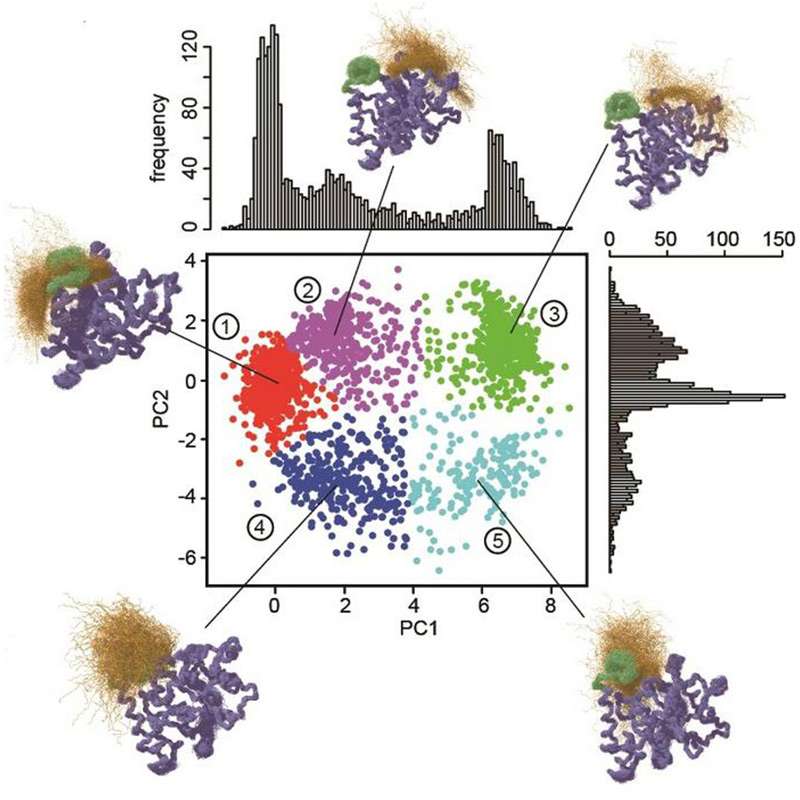
Breakdown of ‘multistate’ nature of YUH1. Analysis reveals that states can be told apart using two parameters (principal components, PC1 and PC2), showing how the gating lid (orange points) can be found at multiple different locations with respect to the crossover loop (green), passing in and out of it over time. Tokyo Metropolitan University. CC BY-NC-ND.
Parkinson’s and Alzheimer’s diseases.
The team demonstrated their method with yeast ubiquitin hydrolase 1 (YUH1), an enzyme in yeast that helps it recycle ubiquitin, a protein that regulates various intracellular events.
YUH1 has an analogue in humans called ubiquitin C-terminal hydrolase (UCHL1), known to be implicated in Parkinson’s and Alzheimer’s diseases.
By integrating multiple analytical methods using NMR spectroscopy, they were able to create an ensemble map for YUH1 for dynamics over the millisecond time scale.
They discovered that two parts of the enzyme near the active part, where proteins are bound, showed strikingly large movements, a ‘crossover loop’ structure and the N-terminus, one end of the protein-capturing structure of the enzyme.
The N-terminus was found to move in and out of the loop, going through a whole range of states before finally capturing a target protein like a lasso before acting as a ‘gating lid’, keeping it in place.
This new mechanism was supported by the way in which mutant versions with incomplete gating lids failed to show the same enzymatic activity.
The team’s findings reveal how the dynamic nature of enzymes plays an important role in how they function.
The method can be applied to a vast array of biological structures in their natural environment, promising a new approach for scientists to access underlying mechanisms and potential pathologies.
The study is published in the Journal of the American Chemical Society.


